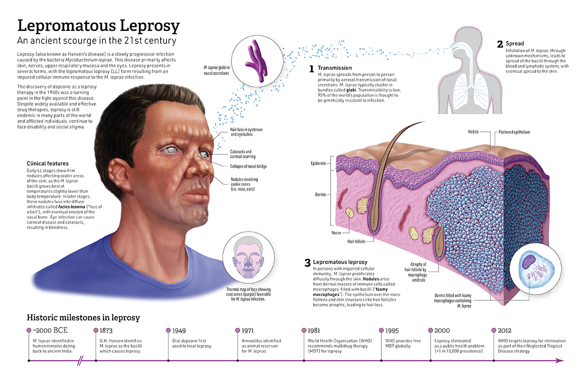
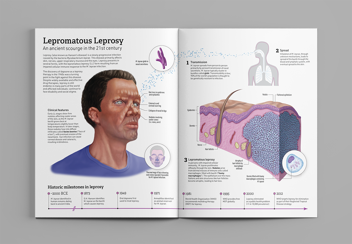
This piece is a mock two-page spread for a popular scientific magazine describing the transmission and histopathology of lepromatous leprosy for an educated lay audience. A head maquette was built in ZBrush, with digital painting of the head completed in Procreate and Photoshop. Vector assets were created in Illustrator and the tissue cube and remaining assets developed in Photoshop. The final composition and graphic design were done in Illustrator.
Audience:
Educated layperson
Date:
December 2019
Supervisor:
Prof. Shelley Wall
Format:
Print
Media:
Adobe Photoshop, Adobe Illustrator, Savage Interactive Procreate, Pixologic ZBrush
References:
1. An, Y., & Zhang, S. (2016). High-resolution, real-time simultaneous 3D surface geometry and temperature measurement. Optics Express, 24(13), 14552.
2. Balamayooran, G., Pena, M., Sharma, R., & Truman, R. W. (2015). The armadillo as an animal model and reservoir host for Mycobacterium leprae. Clinics in Dermatology, 33(1), 108–115.
3. Cardoso, M. de A., de Castro, R. C. F. R., Li An, T., Normando, D., Garib, D. G., & Capelozza Filho, L. (2013). Prevalence of long face pattern in Brazilian individuals of different ethnic backgrounds. Journal of Applied Oral Science, 21(2), 150–156.
4. de Andrade, T. C. P. C., Vieira, B. C., Soares, C. T., Martins, T. Y., Santiago, T. M., & Barreto, J. A. (2017). Lepromatous leprosy simulating rheumatoid arthritis – Report of a neglected disease. Anais Brasileiros de Dermatologia, 92(3), 389–391.
5. Grzybowski, A., Nita, M., & Virmond, M. (2015). Ocular leprosy. Clinics in Dermatology, 33(1), 79–89.
6. Laga, A. C., & Milner Jr, D. A. (2014). Bacterial Diseases. In D. E. Elder, R. Elenitsas, M. Rosenbach, G. F. Murphy, A. I. Rubin, & X. Xu (Eds.), Lever’s Histopathology of the Skin (11th Ed., pp. 658–712). Lippincott Williams & Wilkins.
7. Lory, S. (2014). The Family Mycobacteriaceae. In E. Rosenberg, E. F. DeLong, S. Lory, E. Stackebrandt, & F. Thompson (Eds.), The Prokaryotes: Actinobacteria (4th Ed., pp. 571–575).
8. Massone, C. (2012). Histopathology of the skin. In E. Nunzi & C. Massone (Eds.), Leprosy: A Practical Guide (pp. 115–136).
9. Massone, C., Belachew, W. A., & Schettini, A. (2015). Histopathology of the lepromatous skin biopsy. Clinics in Dermatology, 33(1), 38–45.
10. Nunzi, E. (2012). Clinical Features. In E. Nunzi & C. Massone (Eds.), Leprosy: A Practical Guide (pp. 75–110).
11. Nunzi, E., Massone, C., & Noto, S. (2012). Physical Examination: Skin. In E. Nunzi & C. Massone (Eds.), Leprosy: A Practical Guide (pp. 65–73).
12. Rodrigues, G. A., Qualio, N. P., de Macedo, L. D., Innocentini, L. M. A. R., Ribeiro-Silva, A., Foss, N. T., … Motta, A. C. F. (2017). The oral cavity in leprosy: what clinicians need to know. Oral Diseases, 23(6), 749–756.
13. Salgado, C. G., & Barreto, J. G. (2012). Leonine facies: Lepromatous leprosy. New England Journal of Medicine, 366(15), 1433.
14. Talhari, C., Talhari, S., & Penna, G. O. (2015). Clinical aspects of leprosy. Clinics in Dermatology, 33(1), 26–37.
15. White, C., & Franco-Paredes, C. (2015). Leprosy in the 21st century. Clinical Microbiology Reviews, 28(1), 80–94.
16. Zhu, Y. I., & Stiller, M. J. (2001). Dapsone and sulfones in dermatology: Overview and update. Journal of the American Academy of Dermatology, 45(3), 420–434.


